Why Dental X-Rays Are Necessary
Dental X-rays allow the dentist to see the full picture of oral health, including areas that are invisible to the naked eye.
What X-Rays Detect
Decay between teeth, infections at the tip of a tooth root, bone loss from periodontal disease, impacted teeth, cysts, tumors, and developmental abnormalities are all conditions that X-rays reveal. Detecting these problems before symptoms appear matters because early treatment is less invasive, less expensive, and more likely to preserve the natural tooth.
Supporting Accurate Diagnosis
Clinical symptoms such as pain, swelling, and sensitivity can have multiple causes. X-rays help the dentist pinpoint the exact source and extent of the problem. For example, a toothache may originate from decay, a crack, or an abscess at the root — each requiring a different treatment approach. Accurate diagnosis leads to targeted treatment, which is why X-rays are an essential part of the diagnostic process.
Types of Dental X-Rays
Several types of dental X-rays are used depending on what the dentist needs to evaluate. Each type captures a different view and provides specific diagnostic information.
Bitewing X-Rays
Bitewing images show the crowns of the upper and lower back teeth on a single film. They are the most common type of routine X-ray and are primarily used to detect cavities between teeth and monitor bone levels between teeth. These images are typically taken once a year during routine dental exams.
Periapical X-Rays
A periapical X-ray captures the entire tooth from crown to root tip, including the surrounding bone. This view is used when the dentist suspects an infection at the root, a fracture, or bone loss. Periapical images are frequently taken before root canal therapy or tooth extraction to plan the procedure accurately.
Panoramic X-Rays
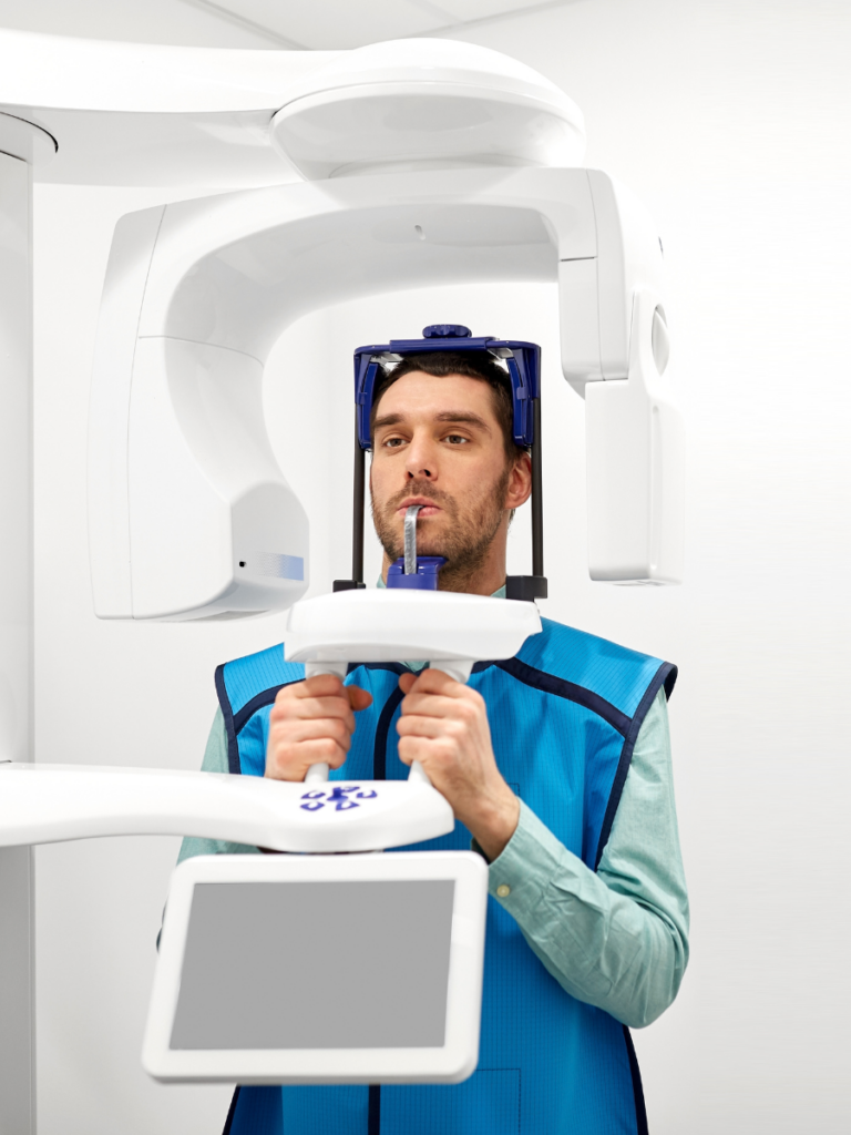
A panoramic X-ray captures a single, wide image of the entire mouth — all teeth, both jaws, the sinuses, and the temporomandibular joints. This view is useful for evaluating wisdom teeth, planning dental implants, assessing overall bone structure, and screening for pathology. Panoramic images are typically taken every three to five years or as part of a new patient exam. The comprehensive nature of this image makes it an efficient screening tool because it reveals conditions across the entire oral and maxillofacial region in a single exposure.
Cone-Beam CT (CBCT) Scans
A CBCT scan produces a three-dimensional image of the teeth, bone, nerve pathways, and soft tissues. This advanced imaging is used for complex treatment planning such as implant placement, oral surgery, and evaluation of jaw joint disorders. The three-dimensional detail allows for precise surgical planning because it shows spatial relationships that two-dimensional images cannot capture.
Digital X-Rays vs. Traditional Film
Modern dental practices use digital X-ray technology, which offers several advantages over traditional film-based systems.
Reduced Radiation Exposure
Digital X-rays use up to 80 percent less radiation than conventional film X-rays. While the radiation dose from traditional dental X-rays was already low, digital technology reduces exposure further. This reduction is especially meaningful for patients who need frequent imaging because cumulative exposure stays well below safety thresholds.
Faster Results and Enhanced Detail
Digital images appear on the screen within seconds, eliminating the wait time associated with developing film. The images can be enlarged, enhanced, and compared side-by-side with previous images, which helps the dentist identify subtle changes over time and share findings with the patient in real time during the appointment. The environmental benefit of eliminating chemical processing is an additional advantage of digital technology.
How Often Dental X-Rays Should Be Taken
The frequency of dental X-rays is based on individual clinical need rather than a fixed schedule for every patient.
Risk-Based Scheduling
Patients with a history of frequent cavities, gum disease, or complex dental work may need X-rays more often. Patients with excellent oral health and low cavity risk may need imaging less frequently. The dentist recommends a schedule based on each individual risk profile, because tailoring the approach ensures the diagnostic benefit justifies the imaging.
New Patient X-Rays
New patients typically receive a full set of X-rays — including bitewings and either a panoramic or complete periapical series — to establish a baseline. These initial images allow the dentist to identify existing conditions, evaluate previous dental work, and create an informed treatment plan.
Are Dental X-Rays Safe
Dental X-rays are among the lowest-radiation diagnostic imaging procedures in medicine. The dose from a single dental X-ray is comparable to the amount of background radiation a person receives in a single day from natural sources.
Protective Measures
Lead aprons and thyroid collars are used to shield the body during X-ray exposure. Digital sensors require minimal radiation to produce a clear image. These precautions are standard practice because they further minimize an already very low level of exposure.
X-Rays During Pregnancy
The American Dental Association confirms that dental X-rays with proper shielding are safe during pregnancy when clinically necessary. However, many practitioners prefer to postpone elective imaging until after delivery unless an urgent diagnostic need arises. The dental team discusses individual circumstances with each patient so that informed decisions can be made together.